
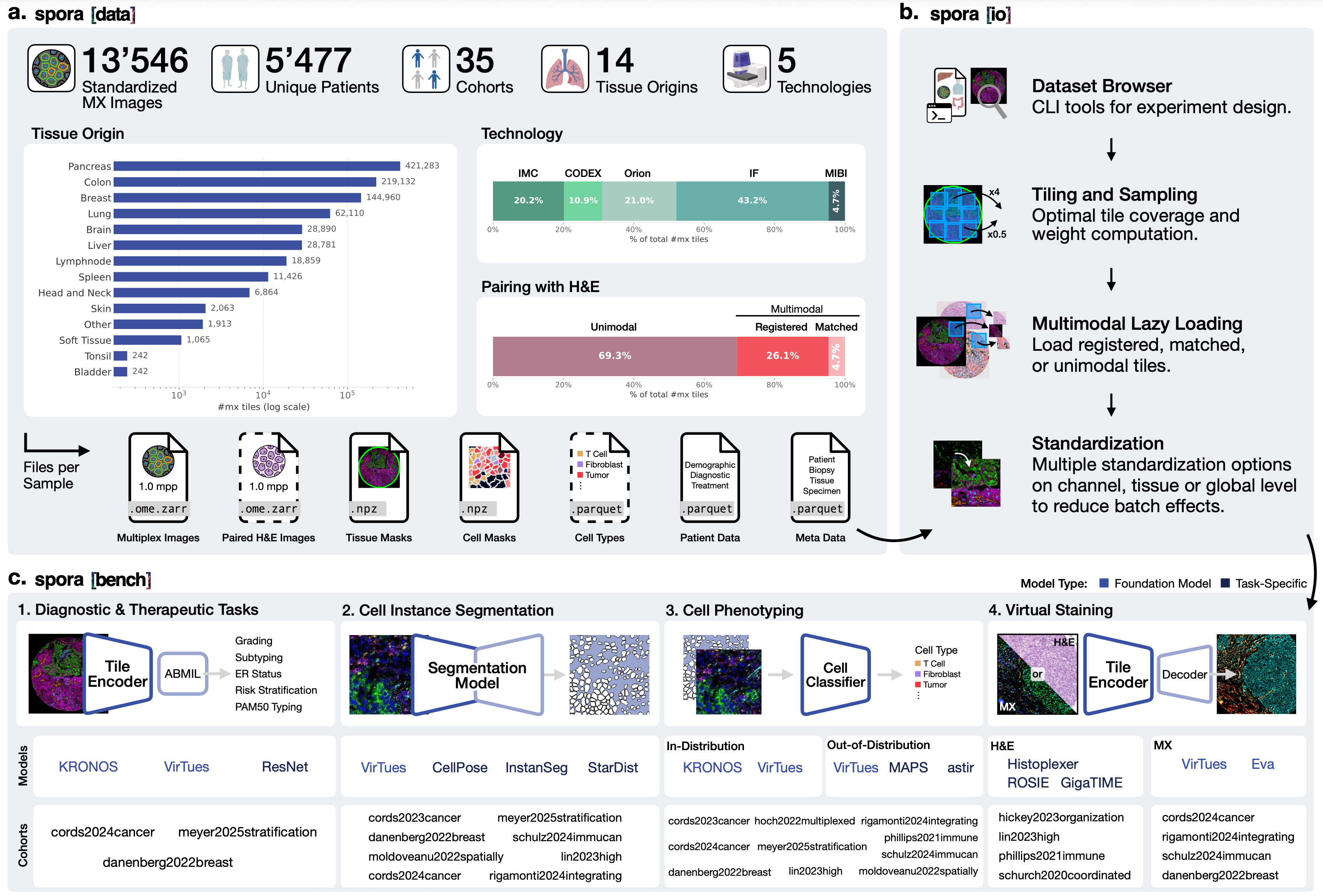
spora. a. spora[data] brings together standardized spatial proteomics cohorts. b. spora[io] provides tools for loading and preprocessing the data. c. spora[bench] supports evaluation of cell- and tissue-level models.
Spatial proteomics measures dozens of proteins in intact tissue sections while preserving the positions of individual cells. It reveals how immune cells, tumor cells, and surrounding tissue interact, helping researchers study disease processes that are difficult to capture with dissociated or bulk assays.
Yet these datasets remain difficult to use together. Studies differ in imaging technology, antibody panels, file formats, segmentation methods, and clinical annotations. Researchers often have to repeat substantial data preparation before they can train a model across cohorts or test whether a biological finding holds up in another study.
We built spora, the spatial proteomics reference atlas, to make that work reusable. It brings together public datasets in a common format, with tools for accessing the collection and integrating new data. The collection is hosted by EMBL-EBI, and our paper describes its curation, software, and evaluation.
A Collection Spanning 35 Cohorts
The current release includes both whole-slide images and tumor cores from tissue microarrays, spanning IMC, CODEX, Orion, MIBI, and immunofluorescence. Assembling the collection took two years of curation with biomedical experts, including harmonizing image formats, reconciling metadata, and reviewing cell segmentations.
Dataset Scale
Modalities and Annotations
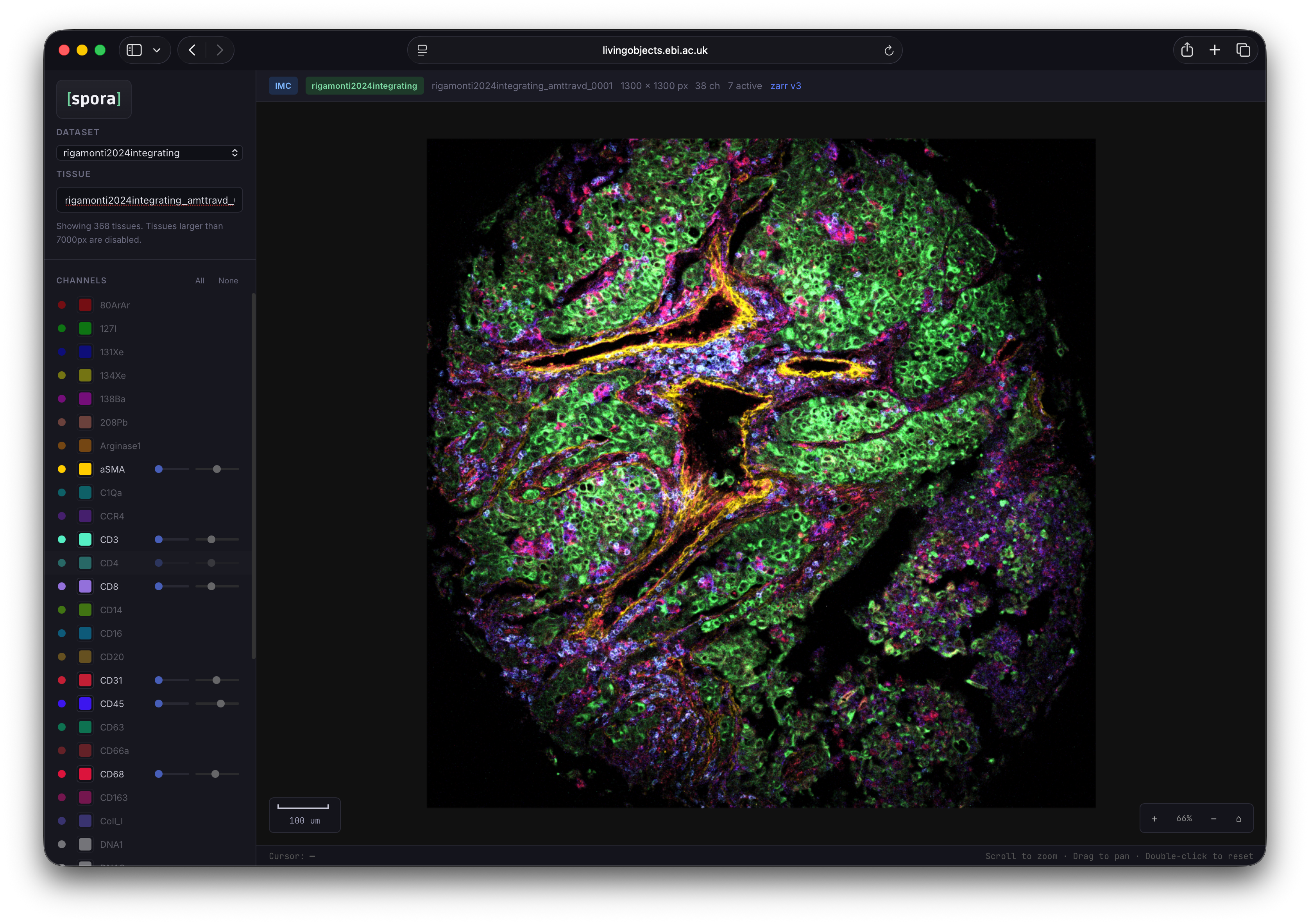
The dataset browser helps researchers identify cohorts with the tissues, markers, and annotations needed for a particular question. The accompanying tissue visualizer supports inspection of individual images and marker channels before downloading the data.

What Is Standardized
spora[data] gives each cohort the same file structure and metadata conventions, with several practical consequences for reuse:
- Images can be loaded consistently. Multiplex images are provided at a common resolution of 1.0 µm per pixel in OME-Zarr, which allows software to read selected image regions without loading an entire slide into memory.
- Channels can be identified across studies. Marker names are standardized and protein targets are linked to UniProt identifiers where applicable. Channel metadata record which markers were measured and which passed quality control. Nuclear stains such as DAPI are identified separately.
- Cell and tissue annotations are available alongside the images. Tissue masks distinguish foreground from background. Standardized cell masks predicted with VirTues accompany the original study masks where available. Sixteen cohorts also include cell phenotype annotations from the source studies.
- Images remain connected to their biological context. Consistent tissue and patient identifiers link samples to structured metadata. Depending on the cohort, annotations include cancer type, grade, stage, treatment, response, survival, and patient demographics.
Where H&E images are available, the metadata distinguish pixel-level registration from matched samples that are not spatially aligned. Registered pairs allow direct comparisons between predicted and measured protein signals at corresponding locations in the tissue.

These conventions make cohorts easier to load and query together. Differences in marker panels, acquisition conditions, and annotation coverage remain, and still need to be considered when designing an analysis.
From Data Access to Model Training
spora[io] provides a Python interface for working with individual cohorts or combining several in one experiment. It loads multiplex images, H&E, and segmentation masks on demand, and supports weighted sampling across cohorts of different sizes. Precomputed intensity statistics make normalization choices explicit and reproducible.
The library also includes tiling that adapts to irregular tissue shapes, improving tissue coverage while limiting background. Together, these tools support parallel model training on high-performance computing systems without requiring each group to build its own loading and preprocessing pipeline.
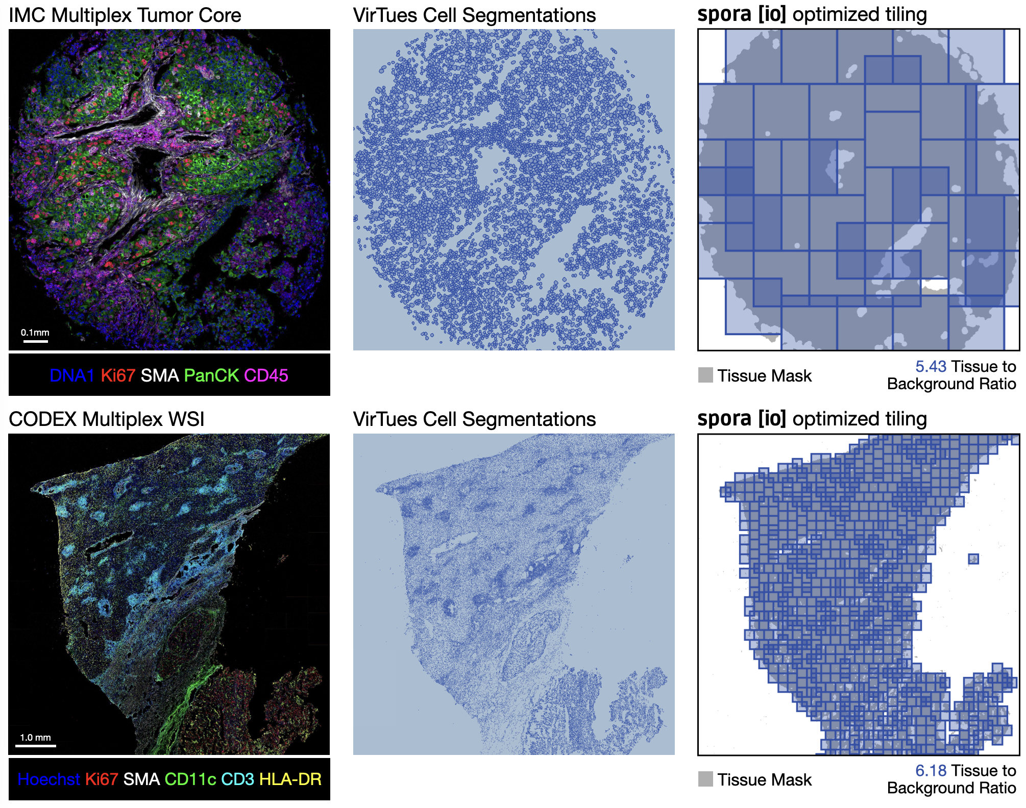
spora[io] tiling. Tiles follow the tissue geometry to improve coverage and reduce background sampling.
Research Across Cohorts
Researchers can use the collection to pretrain foundation models and investigate questions that extend beyond a single study: does a spatial biomarker recur in an independent cohort, can a cell annotation method transfer to another antibody panel, or does a model capture tissue biology across imaging technologies? Paired H&E also supports research on predicting protein signals from routine histology.
To support evaluation across the collection, spora[bench] provides common tasks for cell segmentation, cell phenotyping, diagnostic and therapeutic prediction, and virtual staining. Patient-level train–test splits keep images from each patient in a single split, allowing methods to be compared under consistent protocols.
The virtual staining evaluation illustrates why this matters. Three existing H&E-to-multiplex models showed limited performance on four evaluation cohorts, with correlations between predicted and measured signals near zero for most markers. This finding highlights the value of combining independent datasets with common evaluation procedures to test whether models generalize beyond the studies on which they were trained.
Using and Contributing Datasets
Researchers can also use spora with their own data. Our conversion guide includes an illustrative example how to convert one of the datasets from scratch into the spora[data] format. To assist with that, we provide reusable tools for preparing images and metadata, creating tissue masks, computing normalization statistics, and generating tile coordinates. Once converted, a dataset can be loaded through the same interface as the existing cohorts and analyzed using the methods implemented in spora[bench].
Each cohort remains linked to the study that produced it. Users should cite the original publications as well as spora, and check the applicable dataset licences before reuse: terms differ between cohorts.
The current collection is weighted toward oncology and reflects the tissues, diseases, and technologies represented in available public data. In collaboration with the EMBL-EBI BioImage Archive team, we aim to broaden that coverage through further contributions. If you have questions or feedback, or would like to discuss adding a dataset to the public collection, please contact spora@epfl.ch.
Explore spora
Read the Paper
Dataset curation, methods, and results.
arxiv.org/abs/2610.24002
Use and Extend spora
Data format, downloads, Python tools, and conversion examples.
spora.epfl.ch/docs
Access the Data
Access the dataset collection and visualize tissues images through the EMBL-EBI BioImage Archive.
spora.bioimagearchive.org
This work was led by Benedikt von Querfurth, Johann Wenckstern, Eeshaan Jain, Lukas Klein, Phil Cheng, Yexiang Cheng, Cedric Vincent-Cuaz, Martina Haberecker, Pascal Frossard, Andreas Wicki, Olivier Michielin, and Charlotte Bunne, with collaborators across EPFL, University Hospital Zurich, University of Zurich, University Hospital Geneva, and University of Geneva.
